Knee Effusion – Patellar Dislocation
When a patient has a patellar dislocation, they relate an instability event where they knee “popped out of place.” There is also an effusion.
- Diagnosis
- Apprehension sign
- While pushing down on the medial kneecap, the patient becomes apprehensive and will sometimes try to stop the exam because they think that their kneecap will become dislocated.
- “J” sign
- As the knee is flexed, the kneecap visibly jumps from out of the groove to back into place.
- Treatment
- PRICE
- Physical therapy (PT)
- Surgery
Refer patients to Scottish Rite for continued effusion or recurrent instability.
Knee Effusion – ACL Tear
When a patient describes twisting their knee and it giving out on them or shifting and they have an effusion, they most likely have an ACL tear. Their knee is unstable. There are four ligaments in the knee: the medial knee ligament and the lateral collateral ligament on each side, with the anterior cruciate ligament (ACL) on the front and the posterior cruciate ligament on the back. When the ACL is torn, the knee has more motion, so patients say that their knee slipped or gave out. The best way to check for a torn ACL is the Lachman test.
- The patient lies on their back with their legs out straight and their muscles relaxed, especially their hips and hamstring muscles.
- Bend the patient’s knee slowly and gently to about a 20-degree angle. Physicians may also rotate the patient’s leg so their knee points outward.
- Stabilize the patient’s thigh with one hand and gently move the tibia forward with the other hand.
- If there is a great deal of of motion and instability, it is likely because the ACL is torn
Treatment
- Surgery may be necessary to repair instability or an associated meniscal injury.
Refer any patients with a suspected ACL tear to Scottish Rite.
Knee Effusion – Tibial Spine Fracture
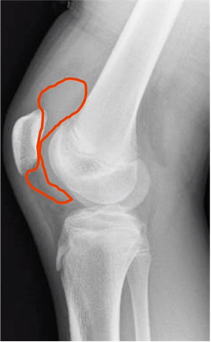
With a tibial spine fracture, the patent usually has a large effusion called a hemarthrosis, or blood in the joint, because of the fracture. These are usually caused by a flexion event like a fall from a bike or skiing or a twist in sport. This fracture will leave a fragment within the “notch” between the thigh bone and the shin bone. This is because instead of the ACL tearing in the middle of the rope, it pulls that piece of bone.
Treatment
- Surgery
- Put the piece of bone back in place
- Casting
- Moving the leg and putting it in a cast may work if it can be placed in a good position
Refer patients to Scottish Rite for immobilization or surgery.
Knee Effusion – Meniscal Tear
It the patient’s reports a twist or pop event and their effusion appears small while experiencing pain on the side of their joint, it is most likely a meniscal tear. Other things to look for to make the diagnosis are focal joint line pain, a loss of extension, a negative Lachman exam, no patellar apprehension, and nothing positive on their X-rays. An MRI may be needed to confirm the diagnosis. The effusion usually means that there is an internal derangement that needs to be treated with surgery.
Conditions with a Chronic Presentation
If the athlete’s injury is a chronic injury, a different set of diagnoses becomes likely:
- Acute vs. Chronic presentation: Chronic
- Stress Fracture
- Has been sore for a while
- Apophysitis
- Patellofemoral Dislocation
- Pain around the kneecap
- Not a specific injury
- Osteochondritis Dissecans
- An idiopathic osteonecrosis below the cartilage surface during development
These conditions generally do not have an effusion, and are all activity-related knee diagnoses.
- Acute vs. Chronic presentation: Chronic
- Effusion vs. No Effusion: No Effusion
- Pain vs. Motion Abnormality: Pain
To determine which condition it is, find out where the pain is located.
- Stress Fracture
- Focal distal femur or proximal tibia
- Tender over a small area around a bone
- Apophysitis
- Focal distal patella or tibial tubercle
- Focally tender
- Patellofemoral Dislocation
- Poorly localized / Not focally tender
- “Horseshoe” sign
- Osteochondritis Dissecans
- Cannot localize
- Deep within
Slipped Capital Femoral Epiphysis (SCFE)
ALWAYS CHECK THE HIP IN ADOLESCENTS WITH KNEE COMPLAINTS
When adolescents have activity related knee pain, often with no inciting event, and display symptoms including a limp, walking with their foot externally rotated and a limited range of motion (especially with internal rotation), it may be SCFE. SCFE is checked with a hip rotational exam. If the patient has equal symmetric range of motion, physicians can rule out SCFE and move on to other diagnoses.
Overuse Conditions – Stress Fracture
Stress fractures are an activity-related pain that often happens after periods of inactivity, like summer. They are associated with high activities like running. Patients are focally tender on their bone, but their knee joints are fine. An X-ray usually shows a stress fracture on their distal femur. The treatment for a stress fracture is forced rest until the patient is pain-free and a gradual return to sports.
Overuse Conditions – Apophysitis
Apophysitis is an activity-related condition with pain focal to only one place. The growth plate is going through a transition with a great deal of stress applied in that area with activities. The two main apophysitis to consider are Osgood-Schlatter Disease in which the patient’s pain is on the tibial tubercle, and Sinding-Larsen Johansson (SLJ) Syndrome in which the pain is on the inferior pole of the patella. The treatments for apophysitis are rest, anti-inflamitories, and quad stretching.
Patellofemoral Pain Syndrome
Unlike Osgood-Schlatter Disease and Sinding-Larsen Johansson (SLJ) Syndrome, the patient cannot pinpoint their pain with Patellofemoral pain syndrome. Patients motion all around the knee in what is called the “Horseshoe” sign. They do not have instability in their knee, but they do have pain around their kneecap. The cause of Patellofemoral pain syndrome is unknown, but it is believed to be related to an abnormal balance of the homeostasis of the muscle strength around the front of the knee. During the exam, physicians determine the “Q” angle, or the quadriceps angle. This is the angle between the quadriceps tendon and the patellar tendon. This angle provides useful information regarding the alignment of the knee joint. “Q” angles greater than 14° are vulnerable to patellar conditions. Physicians also look for poorly developed vastus medialis oblique muscle (VMO), a “J” sign, and pain with patellofemoral compression.
Treatment
- Physical therapy
- Quadriceps strengthening
- Knee balance
- Knee proprioceptive strengthening
- 70% improved with physical therapy, regardless of associated interventions.
Osteochondritis Dissecans (OCD)
OCD is an idiopathic osteonecrosis below the cartilage surface during development. This can lead to cartilage surface cracks, instability and lesion on the joint. OCD can happen with or without trauma. In an X-ray, a radiolucent lesion is visible.
- 2:1 Male to female
- 33% Bilateral
The younger the patient is and the smaller they are, the more likely they are to heal. The location of lesion and the status of articular surface also play a factor in the patient’s healing potential.
Treatment
- Forced rest
- Unloader brace
- Surgery if the patient is older or if the MRI reveals instability.
Meniscal Pathology
A meniscal pathology has a chronic presentation with no effusion, but a motion abnormality instead of pain.
- Acute vs. Chronic presentation: Chronic
- Effusion vs. No Effusion: No Effusion
- Pain vs. Motion Abnormality: Motion Abnormality
The patient says that their knee pops or snaps. They may also have a loss of extension and a limp. These are signs of a discoid meniscus.
Discoid Meniscus
- Discoid meniscus is a congenital malformation of the meniscus
- Affects approximately 1:100 children
- Mechanical symptoms in childhood with no trauma history
- Snapping in the knee usually occurs between the ages of 2 to 6.
- Palpable / audible “snap” at lateral joint line during exam
- Visible bulge at lateral joint line
As children get older, the discoid meniscus presents like a regular meniscal tear. The treatment for this condition is arthroscopic surgery if the patient is symptomatic.
Refer patients to Scottish Rite for mechanical symptoms or loss of motion.

/Ann,-Hip.jpg?width=400&height=266)
