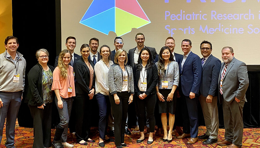
Sports Medicine Team Makes an Impact at Annual Meeting

Sports Medicine Team Makes an Impact at Annual Meeting

Last week, staff from our sports medicine team were in Glendale, AZ for the 7th annual Pediatric Research in Sports Medicine Society (PRiSM) meeting. PRiSM is a unique group of multidisciplinary medical professionals who are devoted to advancing the care for young athletes. The three-day collaborative conference is designed to cultivate relationships among the members and feature advancements in numerous areas of pediatric sports medicine.

With more than ten staff members in attendance, including advanced practice providers, orthopedic surgeons, physicians, physical therapists, biomechanists and research coordinators, Scottish Rite for Children was well-represented throughout the meeting. Selected to present various research projects and serve as moderators, staff had the opportunity to showcase their work and engage in meaningful discussions with other experts in the field. A few of the topics presented included:

Assistant Chief of Staff Philip L. Wilson, M.D., is proud of the team’s involvement. “We have a strong showing at PRiSM each year,” says Wilson. “However, this year, we were represented in almost every session by staff from different departments, which shows our dedication to excellence in every aspect of care for young athletes. PRiSM gives us a great platform to share our knowledge while also giving staff the opportunity to learn from other specialists.”

The team contributed to more than half of the multicenter interest groups who work throughout the year but come together during the annual meeting to brainstorm and discuss the latest findings and progress of projects.

  • Our movement science lab team has made profound progress in establishing protocols to document baseline measurements to aide in projects of interest to the injury prevention group.
  • Sports medicine physician Jane S. Chung, M.D., is a member of the female athlete interest group and the sport specialization group who are both employing surveys to address specific questions.
  • Pediatric orthopedic surgeon Henry B. Ellis, M.D., is the steering committee chair of SCORE – Sports Cohort Outcomes Registry. This effort has already shown very high potential to have major implications in the safety and quality of arthroscopic procedures in youth across the country.
  • Shane M. Miller, M.D., sports medicine physician and concussion expert, is actively involved in a new concussion project that will expand our current understanding and efforts by teaming up with six other pediatric sports medicine programs.

Learn more about the Center for Excellence in Sports Medicine.

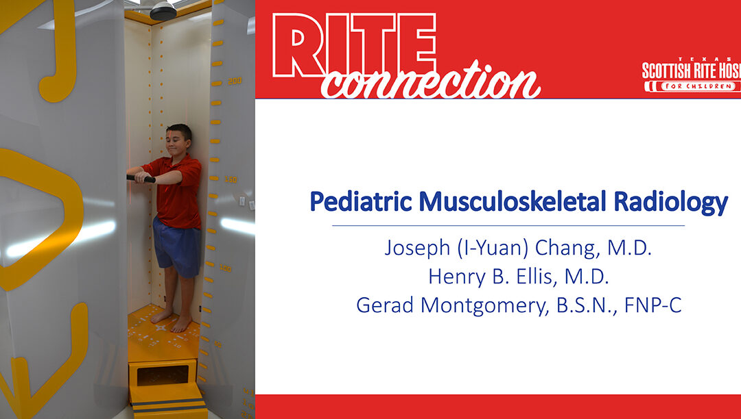
Pediatric Musculoskeletal Radiology

Pediatric Musculoskeletal Radiology

Key messages from Joseph (I-Yuan) Chang, M.D., and a panel discussion by pediatric orthopedic and sports medicine surgeon Henry B. Ellis, M.D., and Gerad Montgomery, B.S.N., FNP-C, at Coffee, Kids and Sports Medicine.

Watch the lecture
Print the PDF

How Advances in Radiographic Imaging Can Protect Patients

Though digital X-rays are the gold standard for many musculoskeletal evaluations, EOS is a relatively new technology designed to achieve results with less radiation. These devices are well-suited for pediatric orthopedics because many treatments, like lower extremity and spine straightening procedures, require periodic imaging to monitor growth and success over time.

Here are several features of how EOS is the best option for some evaluations:

  • Uses very low-dose radiation – uses 1/7 the amount of radiation, compared to traditional X-rays
  • Facilitates accurate assessment of standing alignment – evaluating alignment while a patient is weight-bearing posture provides a more accurate picture of the interaction between the joints of the spine, hips and legs.
  • Creates a single image immediately – with a traditional X-ray, separate films in supine or standing are “stitched” together. This process can be negatively affected by human error (this is done relatively quickly by using computer software at a work station, but may be done incorrectly due to inexperience or carelessness).

IMPORTANT NOTE: It does require a child to stand still for a short period of time, so can only be used when the patient is able to bear weight and can stand still for approximately 10 seconds.

Scottish Rite Hospital has been using the EOS Imaging System since 2016 and had a second system installed with the opening of the Frisco campus in 2018. As pediatric providers, we are committed to using the lowest dosage of radiation possible for studies. EOS has been a useful tool in caring for patients with spinal deformities, lower extremity limb differences and malalignment.

Ordering and Reading Pediatric or Adolescent Elbow X-rays

These tips can be helpful with other X-rays. Watch the full lecture to see how they are applied to ordering and reading an elbow X-ray.

Tips for Ordering X-rays:

  • Always order two perpendicular views – X-rays are 2-dimensional. To evaluate a 3-dimensional object, a bone or joint, two views are necessary. In most cases, the anteroposterior (AP) view and the lateral (LAT) view will suffice.
  • When reading a radiology report, remember that the radiologist does not have the advantage of the complementary physical exam. This is critical to pair with the reading of the imaging. When placing an order, include a note about the clinical exam in the order to provide context for the radiologist.

Tips for Reviewing X-rays:
Joseph Chang, M.D., pediatric musculoskeletal radiologist offered “five easy steps” to reading an X-ray.

  1. Is there a positive ‘fat pad sign’? A fat pad sign, also known as a sail sign, is a sign of a joint effusion. A joint effusion is an imaging finding that is highly predictive of radiographically occult injury in the joint. A pediatric elbow has so much more cartilage than an adult, making certain injuries invisible on radiographs.
  2. Is the alignment normal? In the elbow, assess the anterior humeral line (lateral view) and radiocapitellar line (AP and lateral view). Disruptions to these lines are signs of a fracture or dislocation and need to be treated.
  3. Are the ossification centers normal? Ossification centers have a strict order of appearance and disappearance – if one is missing or out of place, an injury may have occurred. The acronym “CRITOE” can be used to help recall the growth plates in the elbow but knowing to look for them is a good first step. Because growth disturbances can be prevented with proper management, refer to a pediatric orthopedic specialist when you are unsure.
  4. Is there a subtle fracture? Evaluate the metaphysis of the bone. The bony cortex should have a nice, smooth slope. Children have soft and more flexible bone, therefore the bone sometimes buckles instead of breaking. These injuries may appear as a blip on the X-rays.
    • CLINICAL TIP: Be careful not to miss a buckle fracture (also known as torus fracture or incomplete fracture) in your imaging review when a patient has these symptoms.
      • Wrist AND elbow pain
      • Loss of terminal extension and pronation/supination
      • Pain over the radial neck
  5. Did you consider the normal variants? Before you finalize your diagnosis, take a step back and see if what looks abnormal is a normal, developmental appearance in a growing child. Skeletally immature patients may have radiolucent growth centers composed of cartridge and sometimes bone. Secondary ossification centers (i.e. trochlea, lateral epicondyle) can have irregular margins or appear as separate ossicles, mimicking traction stress injury or fractures.

“I think that a practitioner correlating a good clinical exam with the first three steps above will help you identify 90% of elbow injuries and fractures in this population” says Henry B. Ellis, M.D., pediatric orthopedic surgeon.

Joseph (I-Yuan) Chang, M.D., is a radiologist with specialty experience in pediatric musculoskeletal radiology practicing at Scottish Rite for Children Orthopedic and Sports Medicine Center. He completed his training at the University of Cincinnati College of Medicine followed by a residency at Cleveland Clinic Foundation.

The radiology staff at Scottish Rite Hospital participates in interactive, preoperative and postoperative conferences with the pediatric orthopedic specialists. Imaging services include X-ray, EOS, musculoskeletal ultrasound, CT and state-of-the-art MRI capabilities on both campuses. They offer on-demand consultations for our team to support high quality and efficient care.

Hospital Doctors Lead the Research and Education in Children with Clubfeet

Hospital Doctors Lead the Research and Education in Children with Clubfeet

At Scottish Rite Hospital, we are dedicated to providing world-renowned patient care. Our team of experts are able to do so through their steadfast commitment to advancing treatment through research and education. The hospital’s physicians and clinical staff are known worldwide for the research conducted in our Center for Excellence in Foot.

Clubfoot is one of the most common pediatric foot conditions that our experts treat. Through research, our team can study this patient population to better understand the condition and develop innovative treatment plans. With voluntary participation from patients, the team is able to review outcomes after treatment and assess how a child with clubfeet is functioning.

Recently, the hospital published two articles on their latest clubfoot research. Below is what our team is learning:

Functional Outcomes of Patient with Clubfeet at 10-year Follow-up
This research was led by Assistant Chief of Staff Lori A. Karol, M.D., Movement Science Manager Kelly Jeans, M.S., and other staff from the Movement Science Lab.

Summary

Our team analyzed outcomes of patients’ feet who received either the Ponseti serial casting (a form of treatment that places a cast from the thigh to the toes after gentle stretching of the foot) or French physiotherapy (daily stretching, exercise and massage, and taping to slowing move the foot to the correct position), and those who later required subsequent surgery. The Movement Science Lab recorded gait analysis, ankle strength, daily step activity and parent-reported outcomes of patients 10 years after receiving treatment.

What We Learned

  1. Children with clubfeet have less range of motion, movement and power in their ankle in comparison to children with normal feet.
  2. There are minimal differences in gait, parent-reported outcomes and daily activity between feet treated with Ponseti casting or physical therapy.
  3. Feet that did not receive surgery had better ankle power and muscle strength than feet requiring invasive joint surgery.

Clinical Significance

This study supports our efforts to minimize invasive joint surgery when treating a patient with clubfoot. Functional studies such as this continue to help our doctors in developing treatment plans for patients diagnosed with clubfoot.

Read the full article

Non-operative Treatment Outcomes for Patient with Non-Idiopathic Clubfeet
This research was led by Chief Medical Officer B. Stephens “Steve” Richards, M.D., and nurse practitioner Shawne Faulks, M.S.N., R.N., C.N.S.

Summary

Our doctors regularly diagnose and begin treating clubfoot during infancy. Since much of the non-operative treatment takes place before developmental milestones, some patients who were initially thought to have “idiopathic” (no known cause) clubfoot may develop other conditions throughout development rendering a “non-idiopathic” diagnosis. Little is known about the treatment outcomes of this population. Therefore, the purpose of this research is to better understand the clinical outcomes of patients who were later found to have non-idiopathic clubfoot, and how their outcomes compare to patients who have idiopathic clubfoot.

What We Learned

  1. Nearly 1 in 10 infants with idiopathic clubfoot were found to later have non-idiopathic clubfoot due to other orthopedic, neurological or developmental disorders.
  2. Non-idiopathic patients can be expected to respond favorably to non-operative treatment.
  3. Patients later found to be non-idiopathic had a greater chance for clubfoot recurrence that required surgery.

Clinical Significance

This study demonstrates the importance of developmental assessments during a child’s clinical visit. Pediatric orthopedic specialists should be proactive in evaluating more than just clubfeet or other orthopedic disorders during follow-up exams.

Read the full article

Learn more about the research in our Center for Excellence in Foot.

Pediatric Foot Disorders: What Are We Learning From Research?

Pediatric Foot Disorders: What Are We Learning From Research?

Scottish Rite for Children Center for Excellence in Foot includes a multidisciplinary team that is dedicated to advancing the treatment for complex pediatric foot conditions. Directed by Anthony I Riccio, M.D., the center conducts comprehensive research into clubfoot – a congenital disorder in which the child’s foot is severely turned inward and pointed downward. In addition, Dr. Riccio leads the research for complex adolescent foot disorders. Alongside an adult foot and ankle surgeon, Riccio works with a diverse group of specialists to analyze these cases and conducts research to advance how the disorders are treated. 

Prospective Evaluation of Treatment for Clubfoot
Purpose:  To help orthopedic surgeons better understand and treat patients with a diagnosis of clubfoot.  

The research team is collecting data from hospital patients who volunteer to participate in the study. The team is currently evaluating the immediate and long-term outcomes of patients who undergo surgical and non-surgical treatment interventions. Participants are followed until skeletal maturity and are asked to participate in questionnaires, motor and strength tests, gait analyses and pedobarographs (a device that measures foot pressure abnormalities) along the way. Currently, there are over 1,500 patients enrolled in the study. This research will establish a protocol for the collection of relevant clinical and functional outcome measures on our patients with a diagnosis of clubfoot.

The Foot and Ankle Registry
Purpose: To further the understanding of the functional and long-term outcomes of adolescents treated for a foot and/or ankle deformity by creating a prospective registry for patients treated at the hospital.

The research team is currently collecting data on adolescent patients who visit the hospital for any form of foot or ankle deformity. Currently, there are about 300 patients enrolled. Some of the deformities include bunions, flat feet, coalitions and several others of varying severity. If eligible, patients are invited to voluntarily participate in the study activities. These include:

  • Questionnaires
  • Clinical photos in the media department
  • X-rays
  • Gait analysis in the movement science lab 

The data collected will provide the research team the opportunity to evaluate the immediate and long-term clinical, functional and radiographic outcomes of this understudied patient population. By doing this, the foot and ankle experts hope to better define the treatment methods for the varying foot and ankle diagnoses throughout the hospital.

Redefining the Juvenile Bunion
Purpose: To understand the formation of juvenile bunions and if experts should treat them differently from the adult deformity. 

This was a study using the questionnaires, X-rays and pedobarographic data from 32 bunion patients in the Foot and Ankle Registry. The study team collected data from the X-rays through measurements of different angles associated with the foot and correlated them to the completed questionnaires from patients and performance during the pedopbarograph. The study team found that greater angles correlated with skewed pressure distribution of the foot resulting in higher pain and less functionality. Overall, this study will open the door to many more comparative projects to assess the best method of treating juvenile bunions.

Learn more about the research being conducted in the Center for Excellence in Foot.

Doctors Travel to Israel for International Pediatric Orthopedic Conference

Doctors Travel to Israel for International Pediatric Orthopedic Conference

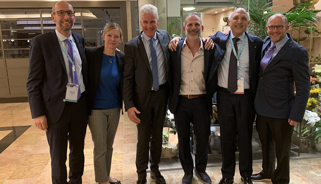
Doctors from the hospital are attending the 38th annual European Paediatric Orthopaedic Society (EPOS) meeting in Tel Aviv, Israel. EPOS is an international association comprised of over 500 surgeons from across the world. This four-day conference allows members to discuss research and collaborate on latest treatment techniques to ultimately advance the quality of care for children with orthopedic conditions.

As active members of EPOS, hospital staff attend and participate throughout this international meeting each year. Several are in attendance, along with past fellows, presenting the hospital’s original research. Below are the projects being presented:

  • Can real time monitoring with a controlled advancement drill decrease plunge depth?
  • Metaphyseal fracture displacement is predictive of intra-articular diastasis in adolescent triplane ankle fractures
  • Gait analysis in children with proximal femoral focal deficiency

In addition to these research presentations, surgeons from Scottish Rite Hospital are directing the first ever web-based joint educational endeavor between EPOS and the Pediatric Orthopaedic Society of North America (POSNA). During this symposium, entitled “Across the Pond: International Differences in Pediatric Orthopaedic Trauma Management,” staff and former fellows from the hospital will discuss and debate current trauma treatment strategies with a panel of European trauma specialists. The debate will be recorded at the EPOS meeting and broadcast worldwide.

Pediatric orthopedic surgeon and director of resident education Anthony I. Riccio, M.D., is one of the hospital’s key representatives for EPOS. “As doctors and leaders within pediatric orthopedics, we have a responsibility to conduct research in order to better understand and find innovative techniques for treating complex conditions,” says Riccio. “It is an honor to travel the world to share our expertise and have the opportunity to both educate and learn from other specialists. Not only does this advance how we care for our patients here in Texas, but it evolves the treatment for pediatric orthopedic patients across the globe.”

Learn more about the hospital’s research.