The Growing Athlete’s Hip: How to Prevent Problems Today and Tomorrow

The Growing Athlete’s Hip: How to Prevent Problems Today and Tomorrow

Download a PDF of this summary.

In this program, our pediatric orthopedic and sports medicine experts described how the skeletal development of the hip is affected by repetitive and extreme movements inherent to athletic activity. The changes, in some cases, can be permanent. Keep reading to learn what we know about preventing irreversible changes and treating symptoms of these sport-related hip conditions.

Apophysitis and Apophyseal Fractures in the Hip and Pelvis

Apophysis is a normal bony outgrowth that arises from secondary ossification centers. The bone fragment will ultimately fuse with the primary bone. The apophysis contributes more to the shape of a bone than the longitudinal growth. Until the ossification center fuses, also referred to as the point at which the “growth plate closes,” the tendon or ligament attached to the apophysis can pull and cause pain in the soft cartilage in the apophysis.

Hip and pelvic apophyses that are vulnerable to acute or overuse injuries are located at the ischial tuberosity, the iliac crest, the anterior superior iliac spine (ASIS) and anterior inferior iliac spine (AIIS). An apophyseal avulsion fracture. An anterior-posterior view of the pelvis is helpful when evaluating complaints in the pelvis so contralateral comparison can be made.
Risk factors for injury includes:

  • Tight muscles and muscle groups
  • Early in the sports season
    • Change in activity from sedentary to active
    • Sudden increase in intensity or duration of training or competition
  • Ignoring activity-related pain
  • Minimal recovery from workouts
    • Year-round training
    • Lack of cross-training
    • Overtraining

Treatment for these conditions is most often nonoperative and is centered around protecting the area involved. Rest, protected weight-bearing, gentle passive ROM and gradual return to play are necessary elements of the plan. Healing and symptom resolution may take 12 weeks or more and radiographic healing is not required prior to returning to sports.

Internal and External Snapping Hip

Athletes may report “popping” in the hip.

If you can see it, it’s likely coxa sultans externus, external snapping hip. This is a condition of the iliotibial band popping over the greater trochanter on the lateral side of the femur. Runners may complain of this when running or walking, and they may describe that it “pops in and out.”

If you can hear it, it’s likely coxa sultans internus, internal snapping hip. This occurs when the iliopsoas muscle, deep in the groin, causes painful popping. This condition is often seen in dancers and tumblers. Treatment includes hip flexor stretching and activity modification.

Femoroacetabular Impingement (FAI)

An overuse injury seen in adolescent and young adult athletes in the hip can be caused by changes in the shape of the femoral head-neck junction (Cam-type) or the acetabulum (Pincer-type). These changes can cause pinching and tearing of the labrum, the soft tissue surrounding the acetabulum that acts to deepen the socket. Early injury from impingement can cause premature hip arthritis. Therefore, this condition is continuing to get more attention with the goal to prevent deformity and consequences.

How does a Cam-type deformity develop?
The femoral head collides prematurely with the acetabulum. The impact causes a change in the shape of the head from being spherical to being more “cam” shaped, or oblong. These may develop secondary to another medical condition in the developing hip, such as:

  1. Slipped capital femoral epiphysis (SCFE) is seen in approximately one in 10,000 may occur and result in avascular necrosis of the femoral head.
  2. Perthes disease – rare condition affecting blood flow in the hip and causes deformity.
  3. Trauma or fracture

In athletes, there is not a primary condition like those listed above. Therefore, idiopathic Cam deformities have been identified in teenage athletes who participate in soccer and other sports. Younger players studied do not show this condition, so the window of opportunity and the exacerbating activity are being studied more closely. Shearing forces may be occurring at the physis to protect the bone, but ultimately may be causing changes in the growth plate and therefore the shape of the femoral head.

Can this be prevented?

Early conversations are looking at the parallel occurrence in the shoulder and elbow in baseball players. Evaluation of the dosage of activity, such as pitch counts in baseball, have been implemented to preserve the anatomy and improve performance in elite athletes. For now, working on proper mechanics and activity modification in adolescence may be our best tools to prevent this deformity.

Considerations and Components of a Hip Injury Prevention Program

Factors that must be considered to prevent hip injuries in adolescent athletes include:

  • Open growth plates
  • Peak height velocity (PHV)
  • High volume of training particularly with loading in rotational and axial movements
  • Sport-specific end range of motion demands
  • Explosive and eccentric demands

Modifiable factors may include:

  • Muscle imbalances
  • Muscle weakness
  • Inflexibility
  • Poor technique
  • Sport-acquired deficiencies
  • Joint instability
  • Overtraining

Five Domains of Injury Prevention Strategies of the Hip

  1. Training Load Management
    Higher incidence of athletic hip pain found with athletes who specialize in a single sport before high school and participate in regular training at earlier ages and four times per week before the age of 12. Recommendations include sampling a variety of sports rather than specializing, monitoring workload, neuromuscular training programs and taking rest breaks from sport (two to three nonconsecutive months/year).
  2. Hip Mobility During Rapid Growth
    Through stretching, dynamic warm-up and eccentric training, hip tissues can stay flexible. Progression of eccentric training can improve the length-tension curve to improve performance and resist injuries.
  3. Motor Control and Stability
    Hypermobility and poor motor control need to be addressed with strategies that improve core stability and teach foundational movement patterns for sport-related movements, such as jumping and landing.
  4. Strength to Improve Imbalances & Specificity
    Once mobility and control are addressed, strengthening can occur. Eccentric adductor & abductor strength can be improved by combining activities, such as the Copenhagen plank and a Nordic Hamstring exercise. Looking for sport-specific strengthening tasks.
  5. Sport-Specific Movement Mechanics
    The culmination of these strategies is executing the sport-specific movement patterns with all of the fundamental movement competence and technical accuracy to ensure safety. Whether the sport demands jumping and landing on a court, changing direction at high speeds on the ice or holding extreme postures on a balance beam, the steps follow a standard pattern.

Implementing Hip Injury Prevention Programs

With confidence that many of these elements are modifiable due to neural plasticity of youth athletes before and during growth, making an effort to prevent injuries is appropriate. Research will continue to define the right and wrong approaches; however, we have some tips that are generally accepted. To avoid detraining, it is recommended to perform activities two to three times per week, approximately 20 min duration, up to 60 min for at least six weeks. It is important to implement it prior to the beginning of a season. Qualified instructors and supervision for continued implementation of the proper techniques are crucial elements of a safe and successful program.

Learn more about hip health in dancers.

This is a summary of a presentation in a monthly series for medical professionals called Coffee, Kids and Sports Medicine. Through events like these, Scottish Rite for Children experts share their experience and knowledge with others to ensure young and growing athletes are getting the best care in every environment.

Five Signs Your Young Athlete May Be Underfueling

Five Signs Your Young Athlete May Be Underfueling

What is underfueling?

When athletes do not eat enough calories (or the right calories) to support their growth and development and all of the training. This may occur periodically due to fluctuations in seasons or training schedules, it is particularly concerning if it happens frequently.

How do I know if my athlete is underfueling?

Without complex calculations, parents can watch an athlete for signs and listen for complaints that indicate underfueling. Below is a short list to help.

  1. The athlete is no longer making improvements in skill and performance or has experienced a sudden decrease in performance.
  2. Injuries take a long time to heal or there are recurring injuries, like stress fractures.
  3. Weight loss that is not otherwise explained. It is important to consider that an athlete may not be losing weight but may still not be getting enough calories.
  4. Delayed growth and development. For a female athlete, an obvious sign is irregular or missed periods or a delay in starting her period.
  5. Frequent dizziness and headaches.
  6. Complaints of constant fatigue.

What should I do if I think my young athlete is underfueling?

  • Make sure he/she is getting three balanced meals a day. Busy teens tend to skip the breakfast meal.
  • Add one or two snacks a day. Often the most reliable snack to add is a bedtime snack. Elite young athletes, especially those struggling with underfueling, typically need at least 3 snacks a day.
  • Increase portions in current meals. Small increases throughout the day can make a difference.
  • Make nutrient-dense swaps in meals and snacks. This way your athlete doesn’t have to worry about adding more food or more eating instances in an already packed schedule.
  • Make drinks count. When athletes need extra calories, include beverages like milk, chocolate milk and calcium-fortified orange juice with meals. Offer sports drinks with practices.

Where can I turn for help if I am concerned?

If your child has lingering or recurrent injuries in sports, delay or changes in menstruation or other concerns that might be related to underfueling, our Sports Medicine team can help. After a medical evaluation with a sports medicine physician, additional services such as a consultation with a certified sports dietitian may be recommended. Call 469-515-7100 to request an appointment.  

Find more resources about sports nutrition for young athletes. 

Five Signs Your Young Athlete May Be Underfueling

Five Signs Your Young Athlete May Be Underfueling

What is underfueling?
When athletes do not eat enough calories (or the right calories) to support their growth and development and all of the training. This may occur periodically due to fluctuations in seasons or training schedules, it is particularly concerning if it happens frequently.

How do I know if my athlete is underfueling?
Without complex calculations, parents can watch an athlete for signs and listen for complaints that indicate underfueling. Below is a short list to help.

  1. The athlete is no longer making improvements in skill and performance or has experienced a sudden decrease in performance.
  2. Injuries take a long time to heal or there are recurring injuries, like stress fractures.
  3. Weight loss that is not otherwise explained. It is important to consider that an athlete may not be losing weight but may still not be getting enough calories.
  4. Delayed growth and development. For a female athlete, an obvious sign is irregular or missed periods or a delay in starting her period.
  5. Frequent dizziness and headaches.
  6. Complaints of constant fatigue.

What should I do if I think my young athlete is underfueling?

  • Make sure he/she is getting three balanced meals a day. Busy teens tend to skip the breakfast meal.
  • Add one or two snacks a day. Often the most reliable snack to add is a bedtime snack. Elite young athletes, especially those struggling with underfueling, typically need at least 3 snacks a day.
  • Increase portions in current meals. Small increases throughout the day can make a difference.
  • Make nutrient-dense swaps in meals and snacks. This way your athlete doesn’t have to worry about adding more food or more eating instances in an already packed schedule.
  • Make drinks count. When athletes need extra calories, include beverages like milk, chocolate milk and calcium-fortified orange juice with meals. Offer sports drinks with practices.

Where can I turn for help if I am concerned?
If your child has lingering or recurrent injuries in sports, delay or changes in menstruation or other concerns that might be related to underfueling, our Sports Medicine team can help. After a medical evaluation with a sports medicine physician, additional services such as a consultation with a certified sports dietitian may be recommended. Call 469-515-7100 to request an appointment.  

Find more resources about sports nutrition for young athletes. 

Osteochondritis Dissecans (OCD) in the Elbow

Osteochondritis Dissecans (OCD) in the Elbow

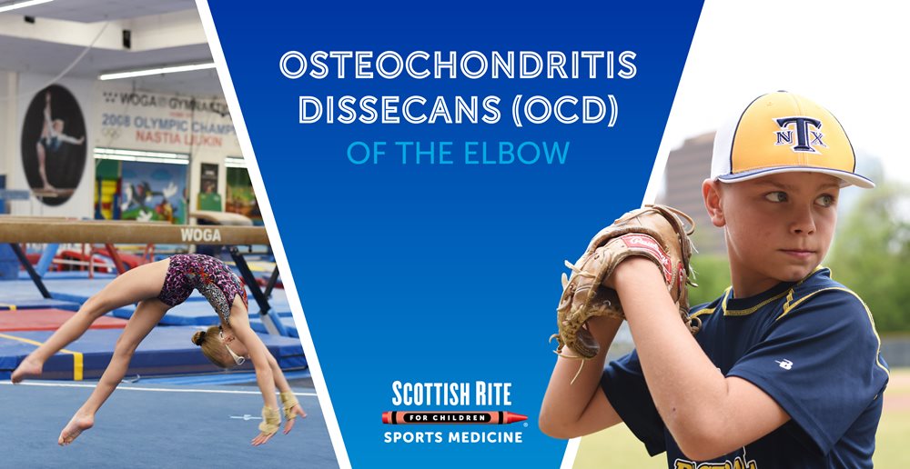
Our Center for Excellence in Sports Medicine treats a wide array of sport-related injuries and conditions in young athletes. One common condition treated is osteochondritis dissecans (OCD) of the elbow. This condition can happen to anyone but is especially common in sports such as gymnastics, tumbling, and baseball.

“This condition often presents to us in very late stages because it develops without symptoms,” says pediatric orthopedic surgeon Philip L. Wilson, M.D. He advises athletes, particularly baseball players and those in weightbearing sports like gymnastics, not to ignore nagging elbow pain. “Painless loss of extension is another sign that should not be ignored,” he says. “Proper diagnosis and early treatment can make a real difference in the course of care and outcomes.”

Our pediatric sports medicine team is a national leader in caring for and studying elbow OCD in young athletes. “The more we learn about the condition and the athletes, the better we can be at treating elbow OCD and teaching others the best way to prevent and manage it,” Wilson says. Here are two examples of Scottish Rite’s work:

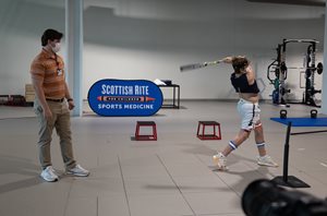
  • An ongoing study called SAFE is open to young athletes, including gymnasts and baseball players. This study is looking at movement mechanics and the causes of injuries in these populations. Check out this video about SAFE testing.

  • study published in 2021, “Elbow Overuse Injuries in Pediatric Female Gymnastic Athletes: Comparative Findings and Outcomes in Radial Head Stress Fractures and Capitellar Osteochondritis Dissecans,” specifically addressed findings in 58 elbows in gymnasts (average 11 years of age) treated at Scottish Rite for Children throughout a course of five years. This study was the first to describe the differences between OCD and radial head stress fractures.

Learn more about OCD of the elbow, its causes, symptoms, treatment, and prevention below.

What is osteochondritis dissecans of the elbow?
The surfaces of the bones inside joints are covered with a smooth, gliding surface called cartilage. Osteochondritis dissecans (OCD) is a condition in which an area of cartilage and the underlying bone begin to soften, crack, or even separate. If left untreated, OCD can cause further damage to the cartilage in the joint and early arthritis.
This is a rare condition that most often affects the knee, but it can also affect the elbow, hip or ankle. In the elbow, the surface on the end of the humerus, the capitellum, is the most affected. This is typically seen in active individuals ages 8 to 19, more often boys than girls.

How does elbow OCD occur?
There are likely several factors, and the exact cause is still unclear. A common cause is a temporary loss in blood supply to an area of bone in a growing child, often combined with repetitive joint impact (overuse). There may be a genetic cause as well. Athletes at risk also often have a history of early sport specialization and year-round training. Some may report a history of a minor injury, but this is likely not the cause of the OCD lesion.

What are the signs and symptoms of OCD in the elbow?
OCD may be present even if there are not symptoms. An asymptomatic OCD lesion, one that does not cause any symptoms, may be identified when evaluating another concern. Signs and symptoms vary and may include:

  • Pain that worsens with activity
  • Popping or clicking
  • Swelling
  • Fluid inside the joint
  • Catching or locking with movement
  • Limited motion

How is elbow OCD diagnosed?
Physical examination, history, and X-rays are used to diagnose OCD in the elbow. Advanced imaging, such as an MRI, is often necessary to fully assess the condition and determine treatment options.

How is elbow OCD treated?
Properly treating and managing osteochondritis dissecans in the elbow lowers the risk of long-term damage to the joint. With diagnosis and treatment in the early stages, tissues may heal with rest and limiting activities that cause pressure on the OCD lesion.

Athletes benefit from continued training while resting their elbows. It is important for our team to help them understand what activities are safe and will not cause further problems on the elbow. Examples of activities to continue while receiving treatment for elbow OCD include:

  • Jogging
  • Stationary bike
  • Core strengthening
  • Lower body weightlifting of resistance training
  • Swimming
  • Golf putting only

These “weightbearing” activities are not allowed because they put pressure directly on the area of the OCD lesion:

  1. Sports of any kind
  2. Handstands
  3. Tumbling
  4. Push-ups, planks
  5. Upper body weightlifting or resistance training

When may surgery for elbow OCD be needed?
Many elbow OCD lesions can improve with conservative, nonoperative treatment. However, surgery may be necessary if the:

  • The OCD lesion appears loose, unstable, or large.
  • Cartilage becomes loose in the joint.
  • Imaging shows an advanced or worsening condition.
  • Symptoms are worsening despite nonsurgical treatment.

What kinds of procedures are used to treat OCD in the elbow?
The choice of surgical procedure depends on the condition of the tissues at the time of surgery. Most procedures are performed using an arthroscope, a camera, and tools inserted through small incisions, but a large surgery may be needed in some cases. Our sports medicine pediatric orthopedic surgeons are experts at treating OCD and can walk you through what to expect.

Procedures that may be offered alone or in combination include:

  • Drilling – drilling holes into the bone to increase blood flow and healing.
  • Stabilizing – inserting a screw, suture, or other piece of hardware to keep loose tissue in place.
  • Grafting – placing biological tissue in the area.

What can be expected after surgery for elbow OCD?
Our sports medicine experts work with every patient to develop an individualized postoperative treatment plan. After surgery, closely following postoperative instructions will protect the joint while the tissue is healing. Exercise and activity recommendations will be different for every patient.

How long does OCD in the elbow last?
Each case is unique, and the timing of returning to normal activity or sports will be discussed with your sports medicine physician, surgeon, or advanced practice provider. Symptoms may last months or years. It’s very important to understand that symptoms may return if the area does not fully recover before returning to repetitive or weight-bearing activities.

How can elbow OCD be prevented?
Overuse injuries like OCD occur with a high volume of training, repetition of certain movements, and early specialization in a sport.

These suggestions can help to prevent elbow OCD and other similar conditions:

  • Learn how to moderate training loads and intensities.
  • Make time for free play and lifetime sports like tennis, golf, cycling, and hiking.
  • Take breaks weekly and between seasons.
  • Learn to properly warm up and perform conditioning for your sport.

Learn more about sport specialization and preventing overuse injuries in young athletes.

Gymnast’s Wrist

Gymnast’s Wrist

Success in gymnastics requires a high volume of training and early specialization. Together, these can take a toll on a young athlete’s growing body. Lindsey Williams, O.T.R., C.H.T., is an occupational therapist who takes care of gymnasts with wrist pain. “I really like working with gymnasts because they are motivated and very compliant with their rest and exercises, but knowing this condition is preventable makes me want to help them catch it before it starts.”

Not too many athletes spend as much time on their hands, so this condition is most common in gymnasts. Because of this, it’s commonly referred to as, “gymnast’s wrist.” Take a few minutes to learn more about this condition and how to recognize early signs and better yet, prevent it.

What is gymnast’s wrist?
Gymnast’s wrist is an overuse injury that causes pain and tenderness in one of the forearm bones, the radius. Distal radial epiphysitis is inflammation in the growth plate near the wrist. This injury is seen, not only in gymnasts, but also in active growing children and teens and is more common in girls than boys. This commonly occurs during periods of rapid growth and/or increased activity.

What causes epiphysitis of the distal radius?
A growth center or epiphyseal plate is an area near the end of long bones that allows for continued growth of a bone. This area is made up of soft cells called cartilage. These weaker cells are at a higher risk of injury.  Repeated stress or compression in this area causes damage and inflammation that can be painful.

Activities that require repetitive weight-bearing through the hands, particularly in extension, include:

  • Tumbling or vaulting
  • Impact or loading in wrist extension with cheerleading and stunting
  • High volume or intensity of training

Treatment is imperative to prevent long-term damage of the wrist. Without treatment, continued trauma to this area can cause the growth plate to become bone (ossify) early which may require surgery in the future to correct. With early and proper treatment, most recover well without surgery.

The initial treatment is rest from impact and weight-bearing activities. A gradual and guided return to normal movement and activities is important.

When pain has improved, an occupational therapist (OT) will guide the progression of exercises, and when cleared by the physician, introduce weight-bearing activities and transition back to sport as strength and pain allow.

How long do symptoms of gymnast’s wrist last?
Untreated, symptoms may persist until completion of growth in this area. In time, stronger bone cells replace the soft cartilage cells, but pain may still come and go for months to years.
To prevent recurrence, it is important to continue the recommended exercises and to avoid excessive training and impact. Pain may come back or worsen during sports or strenuous activities and treatment may be started again. With proper management, most athletes can return to their sport within 3-6 months from the start of treatment.

Can epiphysitis of the distal radius be prevented?
Any athlete that participates in repetitive weight-bearing and loading of the wrist is at risk for this injury.

Some actions to help prevent this include:

  • Warming-up and stretching before participating in weight-bearing activities will reduce stress on joints.
  • Limit or vary physical activities to avoid overtraining and overuse. Spread out training for high-impact activities such as tumbling and vault to separate days and allow a day or two of rest between them.
  • Rest when sore or in pain.
  • Maintain wrist and grip strength to help support the joint and absorb some of the impact.
  • Wear wrist braces such as Tiger Paws® wrist supports to prevent wrist hyperextension and help decrease stress on your wrists.
  • Be aware of changes in wrist pain with increases in training time or when training for a higher level of competition.

 Learn about other overuse injuries in gymnasts.