An Affirmation of Independence

An Affirmation of Independence

Published in Rite Up, 2023 – Issue 1. 

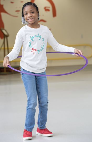
I’m brave. I’m strong. I’m beautiful. I’m perfect — just the way God made me,” says 8-year-old Jordynn, of Crowley, with her mother, DeAdriene. Together, they practice daily affirmations. “Because of Jordynn’s condition, she is different,” DeAdriene says. “When you’re different, it can be a confidence killer. I want her to be the best version of herself that she can be.”
 
Jordynn was born at 30 weeks and spent six weeks in the NICU. At her 12-month appointment, she could not sit up or walk. Her pediatrician said not to worry about walking until she was 18 months old. “At 18 months, Jordynn still wasn’t sitting up,” DeAdriene says. “We’d have to prop her up in a corner.”

A month later, Jordynn was diagnosed with cerebral palsy, a neurological disorder that affects a person’s ability to move. Jordynn’s condition was caused by periventricular leukomalacia, a type of brain injury that is common in very premature babies. In Jordynn’s case, it affected her legs the most. “When we found out, it was really hard,” DeAdriene says. “We were seeing all these doctors, and they said she would never walk independently.”

Jordynn began wearing leg braces, and at age 3, received a gait trainer — an assistive device similar to a walker that supports a child’s mobility. For long distances, she used a wheelchair. As Jordynn grew, she began taking steps on her own, but she could not put her right foot flat on the ground. “The position of her foot was horrible,” DeAdriene says. “Her braces had stopped working because her muscles were so tight.”

DeAdriene had heard about a surgery that she thought could help. When Jordynn was 5, she was referred to Scottish Rite for Children where they sought advice from a team of cerebral palsy experts, including Lane Wimberly, M.D., medical director of movement science and pediatric orthopedic surgeon, and Fabiola I. Reyes, M.D., pediatric physical medicine and rehabilitation physician.

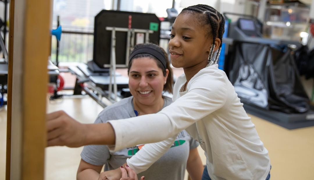
Jordynn underwent a thorough evaluation, including a gait analysis in Scottish Rite’s Movement Science Lab. The analysis measured Jordynn’s walking ability, which aided in designing a customized treatment plan. Through surgery, Dr. Wimberly lengthened her Achilles tendon and hamstring to get her foot into a neutral position. Over the next year, Jordynn regained strength through physical therapy and home exercises. During that time, Dr. Reyes provided tone management by prescribing medication to relieve Jordynn’s muscle spasms, which completely resolved her pain.

“Scottish Rite has exceeded our expectations,” DeAdriene says. “Jordynn is not only walking, she’s running around with her friends. She’s even on the dance team at school!” “Jordynn is happy and active,” Dr. Reyes says, “and that is our ultimate goal for our patients.”

Read the full issue.

Orthopedic Manifestations of Cerebral Palsy

Orthopedic Manifestations of Cerebral Palsy

Content included below was presented at the 2021 Pediatric Orthopedic Education Symposium by pediatric orthopedic surgeon Lane Wimberly, M.D.

Watch the full lecture or download this summary.

At Scottish Rite for Children, we have a multidisciplinary team dedicated to providing excellent care to children with cerebral palsy (CP) through an interdisciplinary approach with evaluation, treatment, and support of the families throughout their childhood. We provide services grounded in evidence-based interventions, employing standardized practices to best demonstrate treatment outcomes for orthopedic and neuro-developmental conditions, including neuromuscular scoliosis, hip subluxation, spasticity, and associated co-morbidities.

With the multidisciplinary approach at Scottish Rite, orthopedic surgery, neurology, pediatric development medicine, movement, orthotics, physical therapy, occupational therapy, science, neurosurgery, psychology, and nutrition experts all work together to determine each patient’s treatment plan.

Cerebral Palsy and Orthopedic Surgery
With this population, it is important to communicate realistic goals and expectations to the patient and family. Surgical recovery may be prolonged—6 to 12 months in some cases – before patients have regained their preoperative strength and functional abilities. Many patients will require new approaches to care and new equipment, like seating systems.

The Scottish Rite utilizes mutual decision making, meaning patients, parents, surgeons, and the rest of the care team work together to make decisions about an appropriate treatment plan for the child, especially when discussing surgery. With these medically complex patients, there are greater risks of surgical complications, which orthopedic surgeons discuss with the patient and the family as part of the decision-making process. Our team helps families cope with the best and less optimal outcomes to ensure the best care for the child.

Gross Motor Function Classification System (GMFCS)
The GMFCS allows physicians to guide treatment and expectations. This standardized tool helps to classify the function of the child on a scale of 1 to 5 depending on their functional level.

  • Level 1 = the child is physically active with a slightly noticeable difference.
  • Level 5 = the child is in a wheelchair and requires assistance with all activities and daily living.

There may be some subtle changes as the child grows and ages, but it is very hard for a child to change one level. Most children achieve their optimum level by age 5 or 6. Children with lesser functional abilities often have a decline in their functional abilities as they age. As the child grows and gets heavier, the inherent weakness with their muscular disorders becomes more apparent, and they may need more assistance.

GMFCS Guide to Surgery
Surgery is optimally offered between 7 and 11 years of age. At this age, recovery is typically easier on the patient and family, and it has shown to be the window to obtain maximum benefit. In addition, contractures at that point are usually becoming less amenable to non-operative treatments.

  • Surgical goals for patients at GMFCS levels 1,2 & 3
    • Maintain function
    • Maintain ambulation
    • Prevent contractures
    • Prevent pain
    • Maybe increase function
      • Not always possible
  • Surgical goals for patients at GMFCS levels 4 & 5
    • Prevent pain
    • Allow ease of care
    • Maintain range of motion
    • Improve sitting tolerance or balance
    • Improve foot positioning
    • Unlikely to improve ambulation
    • May prolong standing tolerance or transfers

Orthopedic and Neuro-developmental Conditions Associated with Cerebral Palsy
Neuromuscular Scoliosis
In periadolescent patients, neuromuscular scoliosis is usually managed with a spinal fusion and implants. The goal of this surgery is to prevent curve progression while improving sitting balance and providing a better seated position.

  • Refer for pediatric orthopedic care if the patient develops:
    • an obvious increase in stiffness of the back.
    • an altered seating posture.
    • a persistent leaning to one side.
    • pelvic asymmetry.

Neuromuscular Hip Dysplasia
Children with cerebral palsy are typically born with normally developed and positioned hips. Over time, excessive linear and rotational muscle forces affect the growth of the femur and pelvis  which may cause the hip to dislocate. The likelihood of neuromuscular hip dysplasia is directly related to the patient’s functional ability – a child with a higher GMFCS level has a higher risk. There is little documented benefit to bracing, Botox injections, or physical therapy for treating neuromuscular hip dysplasia. Surgery is recommended to treat this condition.

  • Early referral and close monitoring can improve surgical outcomes when it becomes necessary. Current guidelines include:
    • Initial assessment at age 2.
    • A supine pelvis X-ray for baseline.
    • Further imaging is based on the patient’s functional level, exam and prior radiographs.
    • Being seen relatively early is most important for non-ambulatory children.

Knee Contractures
Hamstring spasticity can cause knee contractures, which lead to a crouched gait position and challenges with transfers and other care. This can become very taxing as the child moves. Sometimes early muscle releases can prevent or reduce contractures.

  • Refer for pediatric orthopedic care if the patient develops:
    • Asymmetry in knee extension range of motion.
    • Contractures or intolerance to stretching or positioning to prevent knee flexion contractures.
    • Crouched gait or difficulty with ambulation or sitting.

Foot and Ankle Deformities
The foot and ankle are very flexible in children. When they are flexible, braces can be used. Over time, the foot tends to become more stiff, resulting in bony changes that make bracing difficult and less tolerated. Toe walking is the most common orthopedic manifestation of cerebral palsy, due to an Achilles tendon contracture.

When treating foot and ankle deformities, the goal is for the patient to have a flat, braceable, shoable, flexible, and pain-free foot. The goals may differ depending on the age, GMFCS level, and stiffness of the patient.

  • Refer for pediatric orthopedic care if:
    • bracing is not tolerated.
    • contractures develop.
    • foot position is changing.
    • shoe wear difficulties are apparent.

Are you interested in learning more? Visit our on-demand page for more educational opportunities available for medical professionals.
Torsion and Angular Concerns: Treatment Options and When to be Concerned

Torsion and Angular Concerns: Treatment Options and When to be Concerned

This article was recently published in the Pediatric Society of Greater Dallas newsletter. Committed to improving orthopedics care of pediatric patients in all settings, Scottish Rite for Children specialists are regular contributors to this publication for local pediatricians.

Print the PDF

Torsional and angular concerns are common in pediatrics and are often referred to a pediatric orthopedic surgeon for evaluation. An understanding of the normal physiologic changes in typically developing children will provide the practitioner some confidence in discussions with families. While many providers are willing to tell parents the concerns are “physiologic,” this “diagnosis” often leaves the family wondering what is causing the deformities. It is beneficial to demonstrate to parents the source of the difference, discuss the natural history and explain the timeline for improvement.

In-toeing, also known as an internal foot progression angle, is usually caused by metatarsus adductus, tibial torsion and/or femoral anteversion. Typically, metatarsus adductus is seen in infants and is a medial deviation of the midfoot and forefoot on the hindfoot. In most situations, this is a flexible deformity and can be managed by observation alone. Some providers choose to use reverse-last shoes to provide some stretch to the foot, but these are unnecessary in almost all children. More concerning foot positions may be characterized by a deep medial crease, inability to passively push the foot to neutral alignment or a deformity associated with equinus (limited ankle dorsiflexion). If there is worry that the foot position is relatively stiff, referral to a pediatric orthopedic surgeon is reasonable. Once ambulation and shoe wear begin, many of these flexible differences will have resolved.

Physiological internal tibial torsion (Fig. 1) becomes more clinically obvious when children begin walking. A cover-up test on examination (Fig. 3 and 4) will show the proximal tibia is in a neutral alignment with the femur and an obvious bow and rotation of the leg is seen below the knee. The tibial deformity is thought to be due to intrauterine positioning and therefore subtle differences between sides are common. Because of the tibial bow, the child will often appear to be bow-legged (knee varus) as he will widen his stance to reduce foot tripping during walking (Fig. 2). The family can be expected to see spontaneous physiologic improvement by the age of 6-7 years. Be careful when telling families to expect rapid improvement or resolution at preschool ages. Bracing, physical therapy, and other treatments are not indicated in most patients and have never been shown to definitively influence outcome. Should there be obvious asymmetrical torsion or asymmetrical resolution, referral for an evaluation of Blount’s disease would be warranted.

Increased femoral anteversion is normal in infants as physiologic changes are expected in typically developing children. This increased version becomes more clinically relevant as a source of intoeing in the older, preadolescent population. Typical development demonstrates a change in femoral anteversion that continues to improve until early teenage years. In normal femurs, there is 15-20 degrees of femoral anteversion in adults. Femoral anteversion is demonstrated in gait with an internal foot progression angle and families and patients will often notice internally rotated patellae or an ability to sit in the W position. Physical therapy and bracing have not been shown to be effective treatments. Rarely, for teenagers with significant residual femoral anteversion, osteotomies can be performed to improve alignment.

Bowlegged and knock-kneed appearances also may be of concern to families in growing children. In typical growth, children are often born with varus knees (bowlegs) that change to a neutral alignment at approximately 2 years of age and then naturally enter a knock knee (valgus) appearance that can be maximal at 4 years of age. It is advisable to tell families, if the child is seen before 4 years of age, to expect a worsening appearance prior to improvement. A concerned family could take a photograph of the child standing and compare images every six months or so to track changes. Typical adults have approximately seven degrees of valgus and children can be expected to have this appearance by age 7. For these coronal plane differences, asymmetry or failure to demonstrate physiologic improvement are indications to refer to a pediatric orthopedic surgeon.

Physiologic differences in lower extremity alignment are common sources of parental worry. Understanding normal development allows the provider to confidently assuage the concerned parent (and grandparent).