An Approach to Management of Toddler’s Fractures

An Approach to Management of Toddler’s Fractures

Article originally published in latest issue of the Pediatric Society of Greater Dallas newsletter. Written by Gerad Montgomery, M.S.N., FNP-C, and Ray Kleposki, M.S.N., CPNP.

A toddler’s fracture is classified as a tibia shaft fracture in a child age 3 and younger. These patients usually present with either a witnessed or unwitnessed report of low energy trauma to the lower extremity. The mechanism of injury may vary but usually involves some sort of a rotational component. Due to the patient’s age and inability to clearly articulate symptoms or mechanism of injury, these fractures often leave both parents and clinicians anxious and confused. This is confounded by the fact that up to 40% of initial X-rays are negative for obvious bony pathology. To add to the confusion, these fractures usually do not present with swelling or other obvious physical exam findings.

Presentation
The typical patient that presents with this injury is a toddler between the ages of 1 and 3 years with an acute onset of refusal to weight bear or ambulating with a limp.

Three common reports include:

  • History of remote trauma such as a twist and fall.
  • Onset of symptoms after going down a slide with an adult and the patient’s leg getting twisted at the bottom of the slide
  • Unwitnessed incident where patient was playing in another room and eventually found crying on the floor and unwilling to weight bear on the affected extremity.

Regardless of the mechanism, in most cases, the patient will be unable to reliably articulate what caused the injury or where his/her symptoms are arising from. 

Evaluation
When evaluating the patient in this age group, with the presenting complaint of a limp and/or refusal to weight-bear, it is important to consider other conditions that may present in a similar fashion.

Though unlikely, these include:

  • Infection: early presentation of both benign viral infections such as transient synovitis and serious bacterial infections, like osteomyelitis and septic joints, can present with similar initial complaints.
  • Constipation
  • Inflammatory reaction to recent immunizations
  • Chronic or congenital conditions: consider hip dysplasia, cerebral palsy and foot or ankle deformities.

Most of the time, a good history and detailed clinical exam can eliminate dangerous conditions and lead the clinician to an accurate diagnosis. Usually, a step by step exam (starting at the hips and working down to the feet) will reveal pain-free, full range of motion of the hips and knees. This includes classical tenderness to palpation noted over the tibial shaft and pain produced with rotation of the lower leg.  

Treatment
The vast majority of these fractures are stable injuries and treatment is aimed at providing comfort and modifying activities, with the goal of reducing the risk of further injury. Immobilization is appropriate, however a cast is usually not necessary. It has been well documented that a walking boot produces outcomes equivalent to a cast or splint. A removable boot has a lower risk of complications from skin breakdown and higher rate of patient and parent satisfaction. 

Splinting Considerations – When a Boot is Not Available
We frequently see significant skin breakdown after splints or casts used to immobilize patients in this age group. The size of the extremity makes it difficult to properly mold casts/splints in order to prevent friction with skin contact. The most common areas of breakdown are at the back of the heel and anterior crease of the ankle. If you are splinting a patient in this age group, we recommend paying careful attention to the positioning of the foot and ankle and applying extra padding over bony prominences such as the malleoli and at the back of the heel.

The presence of swelling in toddler fractures is minimal and usually not a concern. Therefore, elevation is not a necessary part of the treatment plan, despite the many clinicians and parents who believe that elevation is important for any fracture. While the elevation alone is not harmful, proper elevation techniques should be taught when elevation is recommended.

Proper Elevation – Keep the Heel Off of the Surface
Poor positioning can cause increased pressure leading to skin breakdown. Place a pillow under the calf only, not directly under the knee or heel. Do not assume that immobilization is a benign modality. You must provide the family with warnings and instructions on what to monitor for regarding signs and symptoms of potential complications.

Patient and Family Education
Providing reassurance to the family is a key goal of patient education. These fractures generally heal well with little to no complications in regard to bony healing or future sequela after four to six weeks of immobilization. Additional imaging may be needed in some cases. A child will gradually return to normal activity within a few days of discontinuing activity restrictions and immobilization.
 Figure-1-(1).jpg

Hot Topics in Sports Medicine: Modalities and Trends

Hot Topics in Sports Medicine: Modalities and Trends

Key messages from a presentation by sports medicine physician Jane S. Chung, M.D., at Coffee, Kids and Sports Medicine.

Watch the lecture
Print the PDF

Young athletes present for post-injury care and performance training guidance in many settings from school training rooms to pediatrician offices. All health care providers should be familiar with the basics of popular modalities and trends in order to provide evidence-based advice to children and parents. This article covers four popular areas of sports rehabilitation and performance.

Chung says, “The key message I want providers to hear is that for many of these trends and modalities, most of these studies have been done specifically in the adult population. Evidence and utility for the pediatric population still needs to be thoughtfully investigated.”

Platelet-Rich Plasma: Evidence and Current Applications
Platelet-rich plasma (PRP) is a high concentration of growth factors & cytokines released by platelets to augment the natural healing process. Blood is collected from the patient and processed. The plasma is injected into the treatment area in a clinic or surgical procedure. Some use ultrasound to guide the injection.

Gaps in literature: Standardized volume to inject, frequency for injections, post injection care not determined
Indications: Chronic tendon injuries (tennis elbow, jumper’s knee, Achilles tendonitis), ulnar collateral ligament injuries, rotator cuff injuries, acute muscle injuries and knee osteoarthritis

Show me the evidence: Watch the video to hear a summary of several relevant studies.

Take Home Points

  • There is no evidence, to date, that PRP in acute muscle injuries is superior than placebo or rehabilitation alone.
  • PRP is associated with a reduction in patient reported pain (up to one year) for certain conditions.
  • Despite widespread usage, little is known on benefits of PRP on the musculoskeletal system.

Blood Flow Restriction Technique: What is it, Applications in Therapeutic Setting
Blood Flow Restriction (BFR) technique or training is a form of strength training which is an important component of rehabilitation and performance training. This modality uses partial vascular occlusion while performing exercises at low loads to improve muscle strength, size and endurance.

This technique uses low-load resistance (20-30% 1RM) for training while in the restricted state. This is less than half of traditional heavy-load (60-70% 1RM) strength training. Therefore, this may be appropriate for certain populations where heavy-load training is not appropriate.

How it works: Induce BFR using a pneumatic cuff inflated proximally on a limb. Perform low-load exercise while blood flow is restricted.

Populations where heavy-load strength training is contraindicated and BFR has been studied: Post-ACL reconstruction (ACLR), knee osteoarthritis (OA), adults with sarcopenia and inclusion body myositis.

Show me the evidence:

  • Past decade, research showing BFR in combo w/ LL (light load) training → significant muscle strength and size in healthy individuals
  • Concerns about adverse effects have not been published in studies, only case reports.
  • Promising but not conclusive results for post ACL reconstruction early strengthening and pain for some patellofemoral pain populations.
  • Positive results as an adjunct to traditional physical therapy post-knee arthroscopy.

Take Home Points

  • Clinical applications for BFR training in patients with musculoskeletal conditions are vast.
  • Further studies are needed to study the efficacy and safety of BFR in both operative and non-operative orthopedic conditions.
  • More effective than low-load training alone but less effective than heavy-load training.
  • Limited data is available in the pediatric population.
  • Might be appropriate adjunct therapy for knee OA, patellofemoral pain, post op knee arthroscopy, post-ACLR and muscle injuries (hamstrings).

Whole Body Cryotherapy: What is it, A Cool Trend That Lacks Evidence?
Whole body cryotherapy (WBC) is a brief, full body exposure to dry air at cryogenic temps of -110⁰ to -140⁰ C for two to four minutes, in a nitrogen-cooled cryochamber, where liquid nitrogen fluxes through pipes inside the chamber’s wall.

Gaps in literature: 

  • Lack of standardized protocols for temperature, timing and frequency.
  • Unknown effects on muscle recovery after mechanical overload in athletic populations.
  • Wide variation in study designs.
  • Inability to blind (and unable to eliminate placebo effect).

Take Home Points

  • Possible benefits include enhanced recovery after injuries, post-exercise and counteract inflammatory symptoms from overuse, post-traumatic recovery, pain and performance.
  • NOT FDA regulated, NOT cleared/approved by FDA as a safe and effective device to treat medical conditions.
  • Skilled and trained personnel must control procedures to prevent adverse effects (necrosis, skin burning).
  • Current contraindications: cryoglobulinaemia, cold intolerance, Raynaud’s disease, hypothyroidism, acute respiratory system disorders, cardiovascular disease, purulent-gangrenous cutaneous lesions, sympathetic nervous system neuropathies, cachexia, hypothermia, claustrophobia, mental disorders hindering cooperation during test, pregnant women, children  under 18 (need parental consent).

High Intensity Interval Training: Pros and Cons, is it for Everyone?
High intensity interval training (HIIT) is repeated bouts of high intensity effort followed by varied recovery times. The intense work period can range from five seconds to eight minutes at 80 – 95% of estimated maximal heart rate. Recovery periods can last as long as work periods performed typically at 40-50% of estimated maximal heart rate. Total workout time ranges from 20 – 60 minutes.

Known benefits are consistent with other cardiovascular exercise, these include aerobic and anaerobic fitness, reduced blood pressure, improved cardiovascular health, improved cholesterol profiles, loss of abdominal fat and body weight while maintaining muscle mass, insulin sensitivity and possibly improved brain health.

Contraindication: exertional rhabdomyolysis

Gaps in literature: General lack of studies on the topic, optimal exercise duration and rest intervals remain unclear.

Take Home Points

  • Positive results in studies that include children and adolescents.
  • Living sedentary lifestyle or periods of inactivity, obesity, hypertension, diabetes: obtaining medical clearance from physician may be appropriate prior to starting HITT program.
  • Can easily be modified for people of all fitness levels and special conditions (i.e. overweight, diabetes).
  • Can be performed on all exercise modes: cycle, walk, swim, aqua training or elliptical.
  • Time efficiency: similar benefits as to continuous endurance workouts, but in a less time.
  • Burns more calories especially post workout due to increased excess post-exercise oxygen consumption (EPOC) after HIIT workouts.

Young athletes are highly motivated to return to sport quickly after an injury and will look for any advantage in the process. The highly competitive nature of youth sports is also driving healthy young athletes to seek ways to improve performance. Our responsibility as health care providers is maintain a general knowledge base about treatment options in the market. Understanding the risks and perceived or potential benefits of these and other modalities will help you guide parents and young athletes in making informed choices.

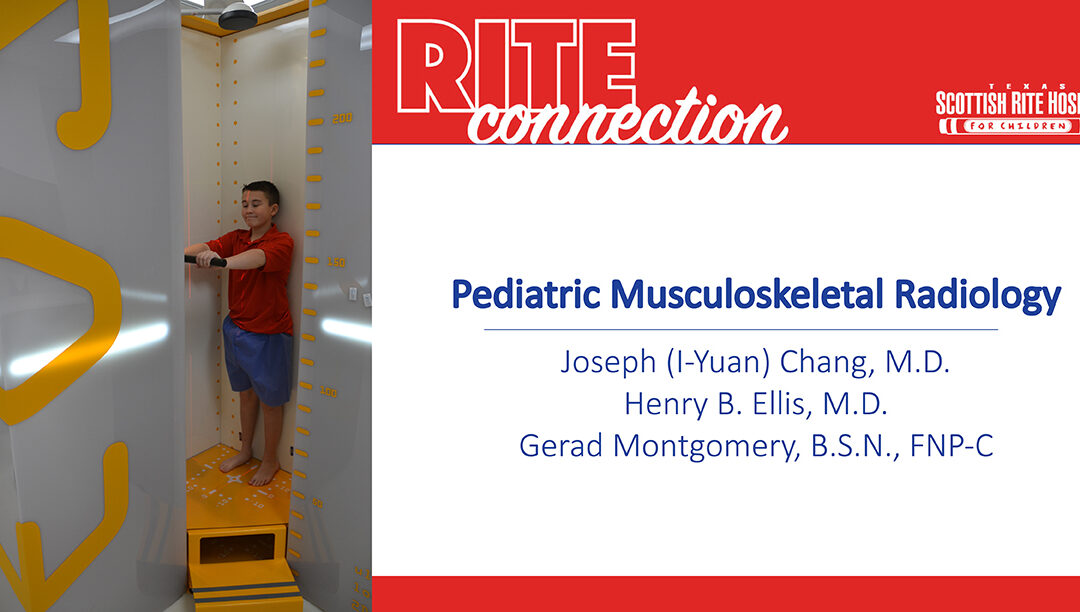
Pediatric Musculoskeletal Radiology

Pediatric Musculoskeletal Radiology

Key messages from Joseph (I-Yuan) Chang, M.D., and a panel discussion by pediatric orthopedic and sports medicine surgeon Henry B. Ellis, M.D., and Gerad Montgomery, B.S.N., FNP-C, at Coffee, Kids and Sports Medicine.

Watch the lecture
Print the PDF

How Advances in Radiographic Imaging Can Protect Patients

Though digital X-rays are the gold standard for many musculoskeletal evaluations, EOS is a relatively new technology designed to achieve results with less radiation. These devices are well-suited for pediatric orthopedics because many treatments, like lower extremity and spine straightening procedures, require periodic imaging to monitor growth and success over time.

Here are several features of how EOS is the best option for some evaluations:

  • Uses very low-dose radiation – uses 1/7 the amount of radiation, compared to traditional X-rays
  • Facilitates accurate assessment of standing alignment – evaluating alignment while a patient is weight-bearing posture provides a more accurate picture of the interaction between the joints of the spine, hips and legs.
  • Creates a single image immediately – with a traditional X-ray, separate films in supine or standing are “stitched” together. This process can be negatively affected by human error (this is done relatively quickly by using computer software at a work station, but may be done incorrectly due to inexperience or carelessness).

IMPORTANT NOTE: It does require a child to stand still for a short period of time, so can only be used when the patient is able to bear weight and can stand still for approximately 10 seconds.

Scottish Rite Hospital has been using the EOS Imaging System since 2016 and had a second system installed with the opening of the Frisco campus in 2018. As pediatric providers, we are committed to using the lowest dosage of radiation possible for studies. EOS has been a useful tool in caring for patients with spinal deformities, lower extremity limb differences and malalignment.

Ordering and Reading Pediatric or Adolescent Elbow X-rays

These tips can be helpful with other X-rays. Watch the full lecture to see how they are applied to ordering and reading an elbow X-ray.

Tips for Ordering X-rays:

  • Always order two perpendicular views – X-rays are 2-dimensional. To evaluate a 3-dimensional object, a bone or joint, two views are necessary. In most cases, the anteroposterior (AP) view and the lateral (LAT) view will suffice.
  • When reading a radiology report, remember that the radiologist does not have the advantage of the complementary physical exam. This is critical to pair with the reading of the imaging. When placing an order, include a note about the clinical exam in the order to provide context for the radiologist.

Tips for Reviewing X-rays:
Joseph Chang, M.D., pediatric musculoskeletal radiologist offered “five easy steps” to reading an X-ray.

  1. Is there a positive ‘fat pad sign’? A fat pad sign, also known as a sail sign, is a sign of a joint effusion. A joint effusion is an imaging finding that is highly predictive of radiographically occult injury in the joint. A pediatric elbow has so much more cartilage than an adult, making certain injuries invisible on radiographs.
  2. Is the alignment normal? In the elbow, assess the anterior humeral line (lateral view) and radiocapitellar line (AP and lateral view). Disruptions to these lines are signs of a fracture or dislocation and need to be treated.
  3. Are the ossification centers normal? Ossification centers have a strict order of appearance and disappearance – if one is missing or out of place, an injury may have occurred. The acronym “CRITOE” can be used to help recall the growth plates in the elbow but knowing to look for them is a good first step. Because growth disturbances can be prevented with proper management, refer to a pediatric orthopedic specialist when you are unsure.
  4. Is there a subtle fracture? Evaluate the metaphysis of the bone. The bony cortex should have a nice, smooth slope. Children have soft and more flexible bone, therefore the bone sometimes buckles instead of breaking. These injuries may appear as a blip on the X-rays.
    • CLINICAL TIP: Be careful not to miss a buckle fracture (also known as torus fracture or incomplete fracture) in your imaging review when a patient has these symptoms.
      • Wrist AND elbow pain
      • Loss of terminal extension and pronation/supination
      • Pain over the radial neck
  5. Did you consider the normal variants? Before you finalize your diagnosis, take a step back and see if what looks abnormal is a normal, developmental appearance in a growing child. Skeletally immature patients may have radiolucent growth centers composed of cartridge and sometimes bone. Secondary ossification centers (i.e. trochlea, lateral epicondyle) can have irregular margins or appear as separate ossicles, mimicking traction stress injury or fractures.

“I think that a practitioner correlating a good clinical exam with the first three steps above will help you identify 90% of elbow injuries and fractures in this population” says Henry B. Ellis, M.D., pediatric orthopedic surgeon.

Joseph (I-Yuan) Chang, M.D., is a radiologist with specialty experience in pediatric musculoskeletal radiology practicing at Scottish Rite for Children Orthopedic and Sports Medicine Center. He completed his training at the University of Cincinnati College of Medicine followed by a residency at Cleveland Clinic Foundation.

The radiology staff at Scottish Rite Hospital participates in interactive, preoperative and postoperative conferences with the pediatric orthopedic specialists. Imaging services include X-ray, EOS, musculoskeletal ultrasound, CT and state-of-the-art MRI capabilities on both campuses. They offer on-demand consultations for our team to support high quality and efficient care.

Prevention of ACL Injury in Our Young Athletes

Prevention of ACL Injury in Our Young Athletes

Article originally published by pediatric orthopedic surgeon Henry B. Ellis, M.D., and Assistant Chief of Staff Philip L. Wilson, M.D., in second quarter, 2019 issue of the Pediatric Society of Greater Dallas newsletter. Key messages were also presented by Philip L. Wilson, M.D., at Coffee, Kids and Sports Medicine. 

Print the PDF
Read the original research

Sports and ACL Injuries: Epidemics in Pediatrics
The dramatic increase in anterior cruciate ligament (ACL) injuries, particularly in female athletes, and an increase in surgeries in young athletes requires attention by all of us. Our research has demonstrated almost a two-fold increase in ACL reconstruction performed in female athletes since 2009. In a large epidemiology study reported by the American Academy of Pediatrics (AAP) in 2014, female soccer, basketball and gymnastics are among the top sports with highest ACL injuries rates. Male football is also considered a high-risk sport for ACL injuries.

Although there are obvious benefits of youth sport cultures, annual increases in participation rates of children and adolescents in organized and year-round sports now allows for more opportunities for injuries to occur. Youth that play soccer or other sports that require pivoting may be especially vulnerable. Although most of us want to see our kids (and patients) win, a healthy and safe attitude towards organized sports is wise.

How do ACL Tears Occur?
The ACL, which is located in the center of the knee, serves to limit rotation and forward movement of the tibia. When overloaded in a vulnerable position, without the support of coordinated muscles, the ACL (with linear collagen similar to a rope) tears or stretches when stressed beyond capacity. Often, this occurs when stopping suddenly, landing with an off-centered pelvic position, cutting or twisting movements or with a direct hit to the knee. Youth might feel a painful pop within the knee, experience their knee giving way and observe swelling when an ACL tear occurs.

The knee is often unstable with a torn ACL. Participating in sports with an unstable knee may lead to damage of other soft tissues (menisci and articular cartilage) in the knee. Therefore, surgery may be necessary.

ACL Injury Treatment
An ACL injury can often be successfully treated. An athlete who wishes to return to sports that involve jumping, cutting and pivoting, may require surgery to re-build the ligament. Even though almost all kids will return to sports, surgery in a young athlete can be stressful, traumatic and may take up to one year of rehabilitation. Although risk to the growth plate exists during this surgery, orthopedic specialists who are trained in pediatric orthopedics and routinely treat growing children with these injuries can minimize these risks to a negligible incidence.

Pediatric Sports Injury Prevention Programs
ACL Injuries and sports-related knee injuries are preventable. This has been established in numerous studies. The risk of a non-contact ACL injury may be reduced if the muscles of the central and lower body are strong and well-coordinated. Programs centered around coordination and balance, strengthening and falling techniques can improve biomechanics and help prevent injury. While there is a role for “old school” warm-ups, such as running, research has confirmed that training to improve the efficient and timely contraction of muscles to stabilize the knee (neuromuscular control) may reduce ACL injury or re-injury.

What Can Pediatricians Do to Evaluate for ACL Injury Risk? 
While evaluating an athlete’s safety to participate or return to high-risk sports following an ACL injury, the pediatric sports medicine community often employs balance and strength testing. Single leg squat endurance while maintaining proper in-line knee form, drop-landing knee form, and ability to perform single leg balance maneuvers at an appropriate age and symmetry level (Y balance testing -YBT) are common measures for knee safety evaluation.

Pediatricians may consider simple screening techniques to establish high risk patients or in those who play high risk sports. Some techniques include a single leg balance assessment or a standing double or single leg squat in the office, while observing for diminishing in-line knee control (see photo). Another examination of core strength that may also be useful is to have the patient hold a plank for 60 seconds. This might best serve as a conversation starter for families with pubescents and adolescents in pivoting sports, such as soccer or basketball. A “when in doubt – refer for knee injury prevention training” strategy may be safest in this high-risk population.

The following training tips may also reduce the risk of an ACL injury:

  • Encourage kids to play for fun first, and then play to win.
  • Recommend variation in their sports throughout the year. This will help maintain long term athletic development and minimize overuse injuries.
  • Train for activity, but be cautious of fatigue that may develop during year-round single sport play.
  • Perform core body, hip and thigh strengthening exercises.
  • Practice wide-based, flexed-knee squatting, jumping and landing techniques.

Education to families and youth athletes regarding these injury prevention strategies and programs is an important wellness initiative for all of us who care for these children. Contacting a community pediatric sports medicine partner is an effective way to gain more information and access to available programs. As participation in youth sports continues to rise exponentially, all of us caring for these children will continue to be challenged to provide valuable and timely “ounces of prevention” for their knees.

Sports Preparticipation Physical Evaluations: Why Are They Important?

Sports Preparticipation Physical Evaluations: Why Are They Important?

Overview of presentation from a Coffee, Kids and Sports Medicine lecture. 

Print the PDF

Every year, almost 60 million children and adolescents run onto their respective fields, courts and rinks to participate in organized sports in the United States. To ensure the safety of these young athletes, almost all states require some level of sports Preparticipation Physical Evaluation (PPE), which is meant to screen athletes for injuries, illnesses or factors that may put them or others at risk.

“The primary objective is to screen for conditions that may be life-threatening, disabling or may predispose to injury or illness,” says Shane M. Miller, M.D., a sports medicine physician at Scottish Rite for Children Orthopedic and Sports Medicine Center in Frisco. “A PPE can also serve as a valuable health care entry point for many young athletes and help the physician determine the general health of the child.”

Miller says an extremely important aspect of a PPE is a thorough medical history for the athlete and his or her family. An accurate medical history can be instrumental in detecting 88% of medical conditions and 67% of musculoskeletal conditions. Without a reliable medical history, the source of an issue may be more difficult to identify. It is estimated that less than 40% of forms completed by an athlete correlate with those completed by their parents. Therefore, the parent or guardian should contribute to the history every time.

Questions to enhance a medical history discussion during the PPE:

  • Have you ever had an injury to a bone, muscle, ligament or tendon that caused you to miss a practice or a game?
  • Have you ever had a head injury or concussion?
  • Has anyone in your family died of heart problems or passed away suddenly before age 50?
  • Have you or anyone in your family had a heart condition diagnosed?
  • Have you ever passed out or nearly passed out DURING or AFTER exercise?
  • Have you ever had discomfort, pain, tightness or pressure in your chest, or does your heart ever race or skip beats (irregular beats) during exercise?

Many organizations and schools require a PPE or health clearance for youth formally participating in sports. Since the intent is to enhance the safety of sports participation, it should become an integral part of the health screening exams for any active patient. Miller reports that a very small number of athletes are denied participation from their sport following a PPE.

“The PPE is a screening tool based on the principles of prevention and education,” Miller says. “It is okay to require further evaluation prior to clearance—that evaluation can establish the foundation for a trusting relationship between physician and athlete that can help keep the athlete healthy and safe throughout their athletic career.” In fact, 3-13% of athletes will require further evaluation.

Common reasons for further evaluation:

  • Chronic ankle sprains or other previous injury
  • Signs or symptoms of overuse injury
  • Recent or multiple concussions
  • Concerns of under fueling for sports participation
  • Recurrent stress injuries or fractures
  • Cardiac – concerning heart murmur, symptoms or family history

The fifth edition of the American Academy of Pediatrics’ text on Preparticipation Physical Evaluation is available now on the AAP website. The fifth edition includes new chapters on transgender athletes, female athletes, mental health and incorporating PPE into routine health supervision care.